
High disease burden
A 26% prevalence of cardiovascular conditions among individuals aged 40 and above, and 7% among young adults aged 25–40.
Serving man is serving god · Vivekanand Vaidyak Pratishthan
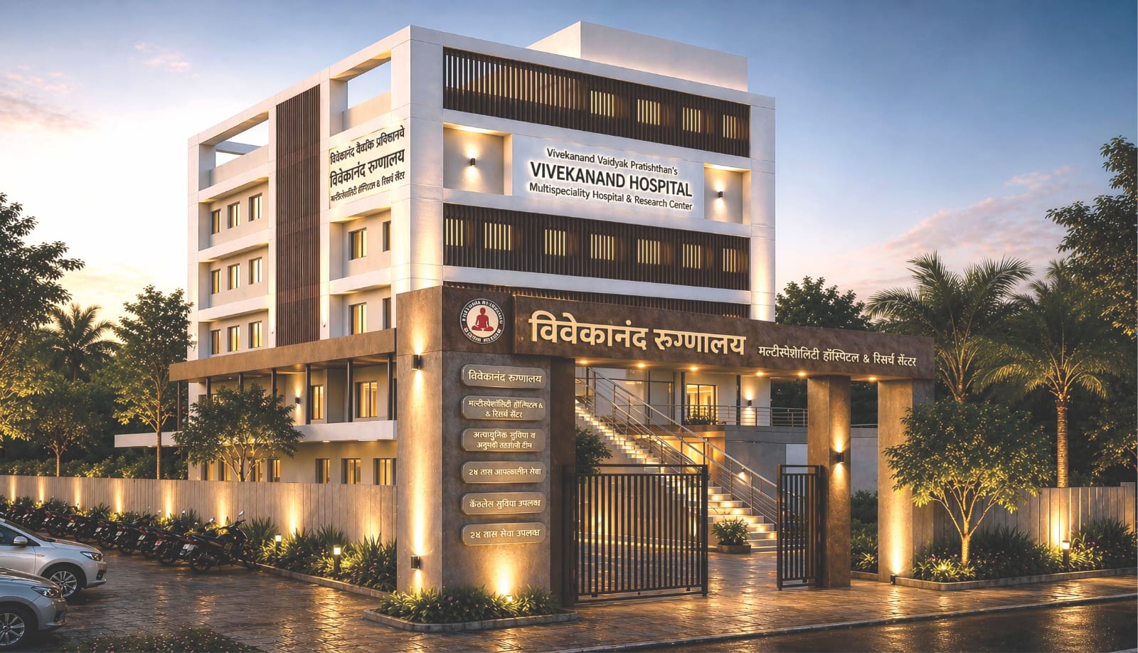
Cardiac care expansion
Bridging the cardiac care divide — REC Ltd. CSR grant to establish a state-of-the-art Cath Lab at Vivekanand Hospital, Bamnoli (Kupwad), Sangli.
Announcement
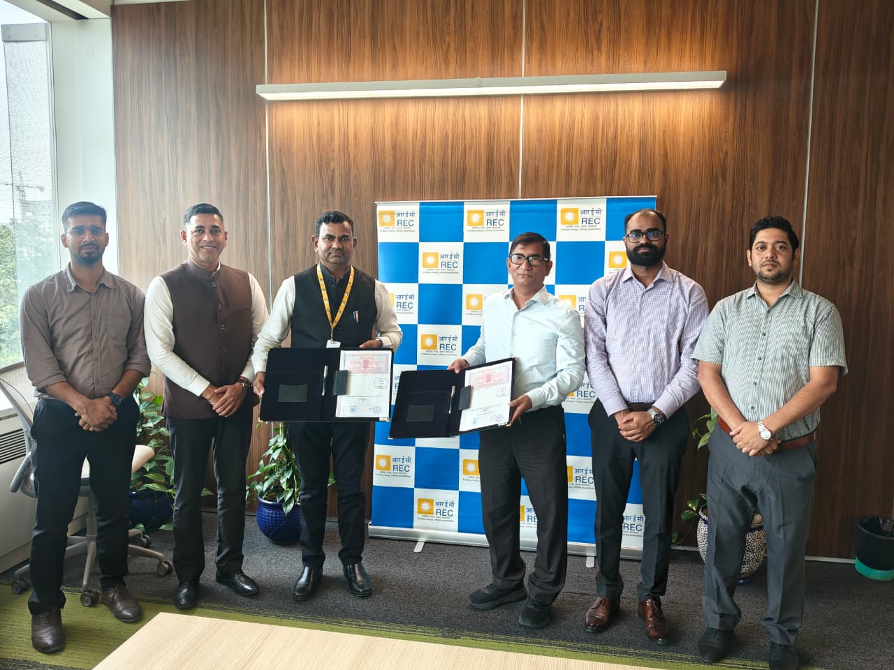
Vivekanand Vaidyak Pratishthan (VVP) is proud to announce the receipt of a major Corporate Social Responsibility (CSR) grant from REC Ltd. This generous support will fund the establishment of a state-of-the-art Cardiac Catheterization Laboratory (Cath Lab) at Vivekanand Hospital (Charitable Multispecialty Hospital & Research Centre) in Bamnoli (Kupwad), Sangli.
This milestone project will bring critical, life-saving cardiovascular and interventional neurovascular care to underserved rural and semi-urban communities across Western Maharashtra and northern Karnataka.
Vivekanand Hospital (Charitable Multispecialty Hospital & Research Centre)
A/P: Bamnoli (Kupwad), Taluka Miraj, District Sangli,
Maharashtra, India — 416 436
Need assessment
Cardiovascular diseases (CVDs) and strokes have surged across both rural and semi-urban populations. In Sangli and the surrounding districts including Kolhapur, Satara, Solapur, Belagavi, and Vijayapura, most families depend on agrarian labor, small-scale industries, and informal employment. A community health survey conducted across 2,200 households (a total 12,742 individuals) in Sangli district revealed stark realities:
A 26% prevalence of cardiovascular conditions among individuals aged 40 and above, and 7% among young adults aged 25–40.
In Sangli district, no government hospital and no charitable facility currently operates a cardiac Cath Lab. Advanced facilities exist exclusively in private hospitals.
When acute emergencies like heart attacks or strokes occur, rural families face devastating choices. Long transit times to distant facilities compromise the vital "golden hour," and the steep costs of private interventional care often push economically vulnerable households into deep debt and poverty.
The solution
With the vital support of REC Limited, Vivekanand Hospital is bridging this critical healthcare gap by establishing a full-fledged, multi-purpose Cath Lab designed to serve the community for over a decade. The new facility will provide round-the-clock emergency and elective interventions either free or at deeply subsidized rates, directly benefiting more than 1,500 underprivileged patients every year.
Clinical capability
Diagnostic coronary angiography, balloon angioplasty, stenting, atherectomy, and heart valve interventions.
Pacemaker and ICD (Implantable Cardioverter-Defibrillator) implantations, cardiac ablation, and congenital heart defect closures.
Emergency mechanical thrombectomy for ischemic stroke, cerebral angiography, and treatment of brain aneurysms and vascular malformations.
Diagnostic catheterization and peripheral angioplasty/stenting.
Integrated real-time radiation dose monitoring compliant with Atomic Energy Regulatory Board (AERB) and BARC standards to protect both patients and clinicians.
Digital road-mapping and real-time plaque quantification for precise intervention.
Supported by uninterrupted 3-phase power, 30-minute full-load backup UPS, and dedicated intensive coronary care infrastructure.
Implementation
To ensure high clinical standards and swift execution, VVP is executing a phased commissioning plan:
Finalizing the specialized cleanroom infrastructure, HVAC, radiation shielding, and integration with the hospital information and PACS digital imaging networks.
Deploying specialized cardiac imaging hardware, onboarding dedicated full-time cardiologists, interventional radiologists, cath lab technicians, and specialized nurses.
Completing AERB and state regulatory licensures alongside rigorous infection control and emergency workflow SOPs.
Beginning with an operational soft launch followed by a full-scale opening for 24/7 cardiac emergency admissions.
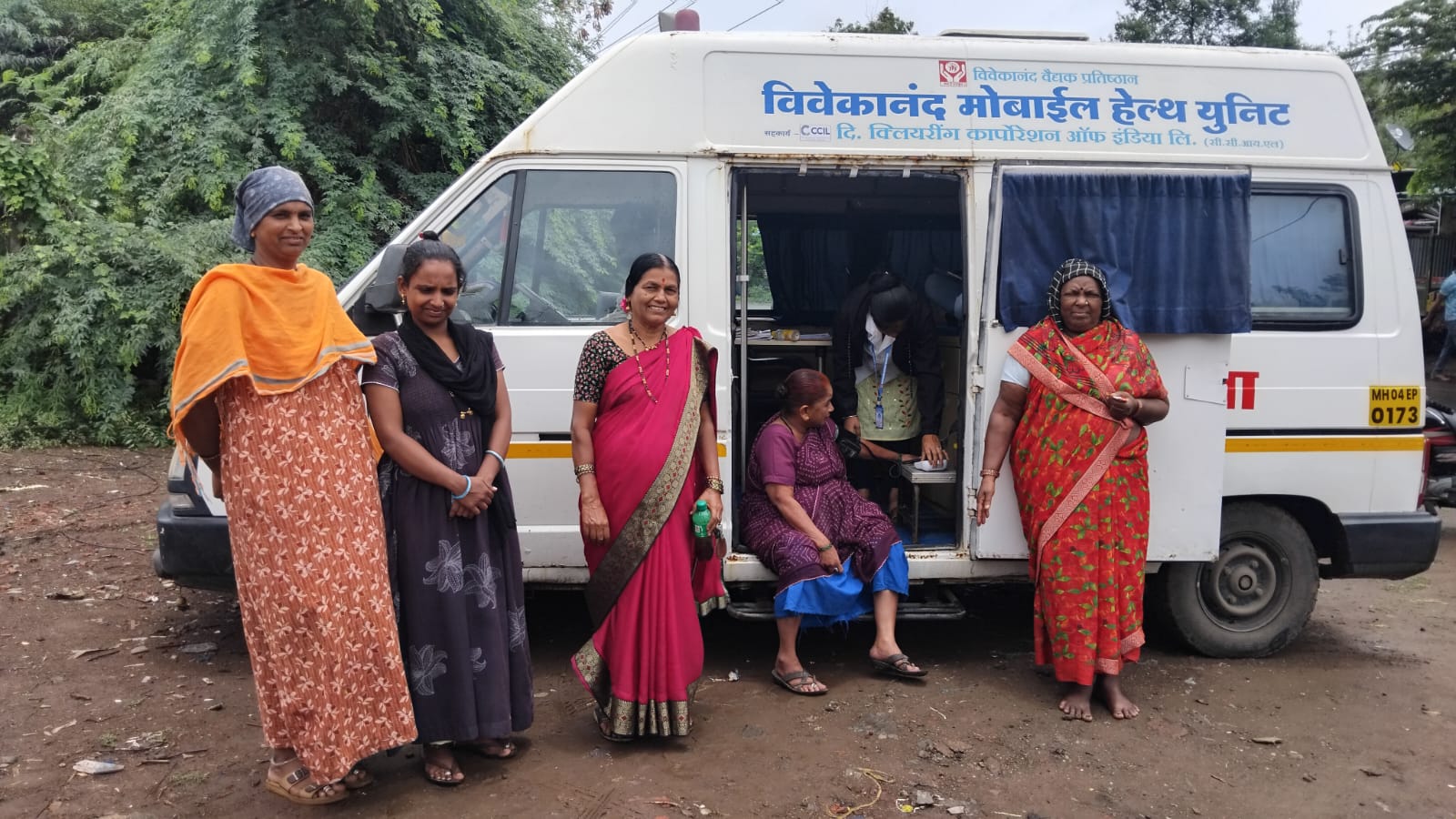
Beyond the initial equipment commissioning, VVP is establishing a comprehensive community referral network connecting Primary Health Centers (PHCs) and rural clinics for rapid diagnosis and timely referrals. Through tiered subsidized pricing for non-indigent cases, local donor backing, and preventive heart-health awareness camps, the hospital will ensure that the unit remains self-sustaining, community-driven, and permanently accessible to those who need it most.
Vivekanand Vaidyak Pratishthan extends its heartfelt gratitude to the leadership and CSR committee of REC Limited. By investing in critical healthcare infrastructure for rural Maharashtra, REC is safeguarding thousands of families from preventable mortality and economic distress. Together, we are transforming rural healthcare to deliver timely, dignified, and advanced cardiac care to the last mile.
From the project brief
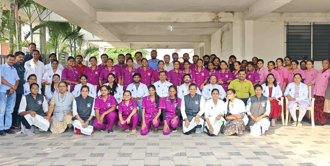
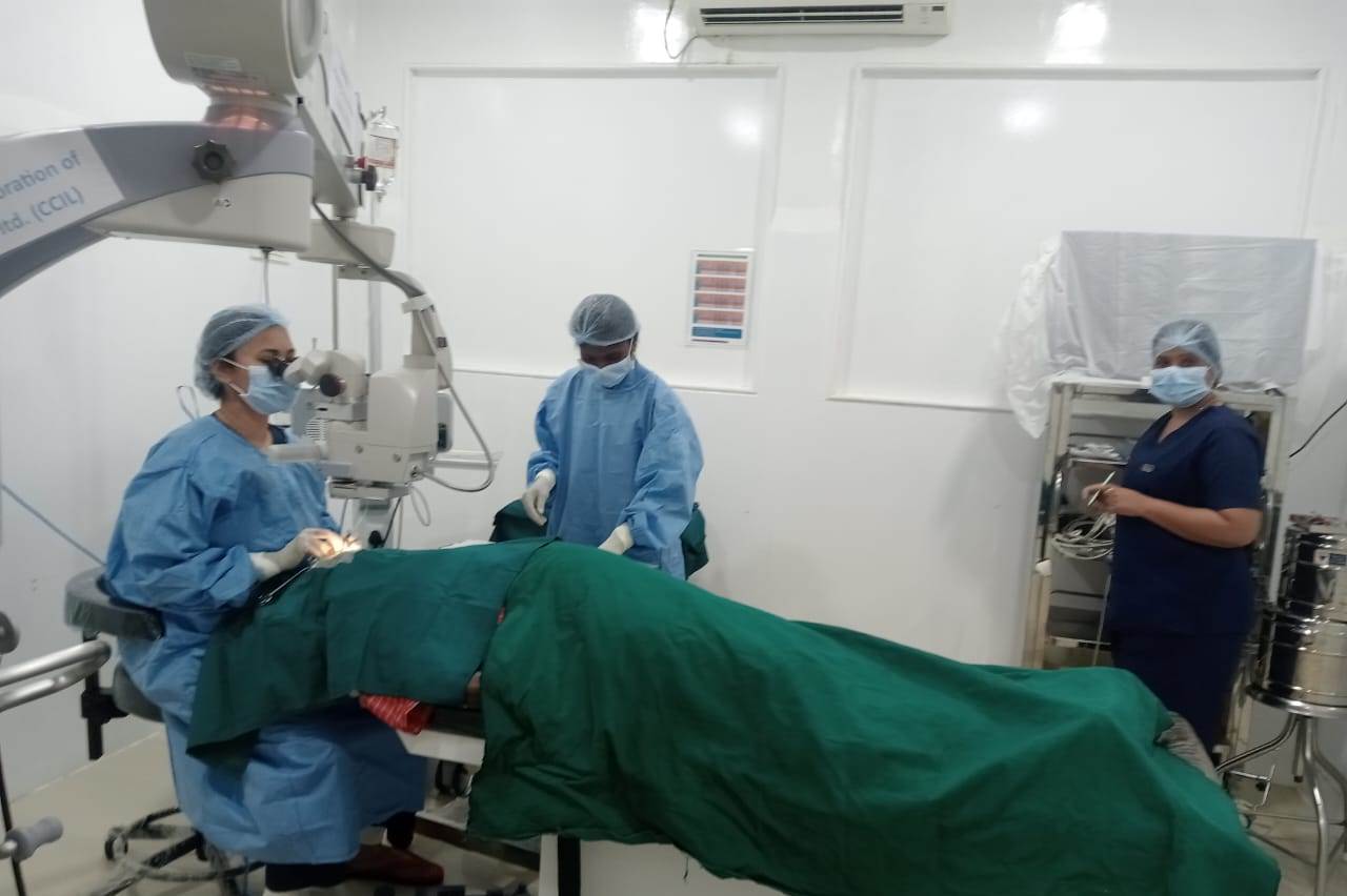
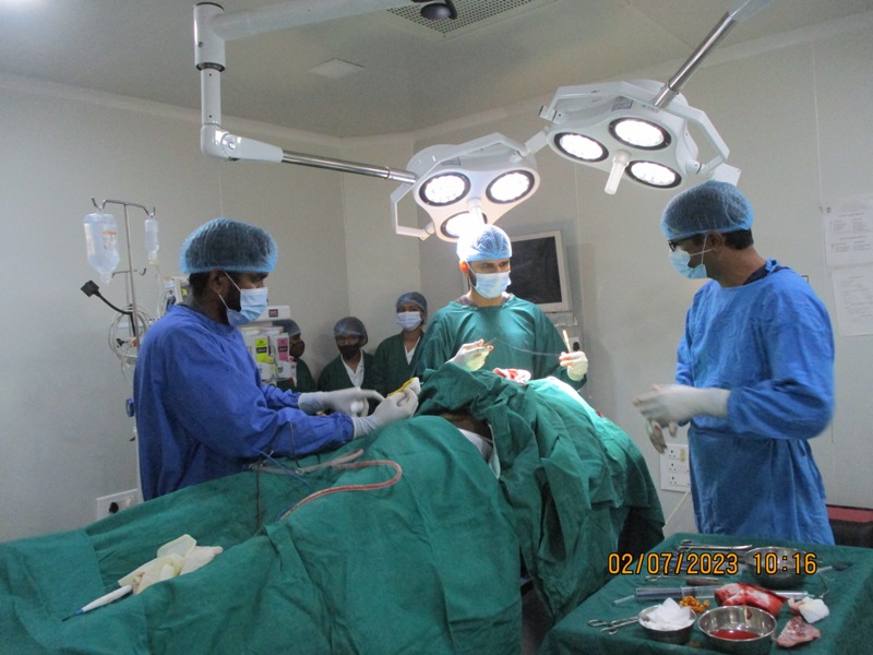
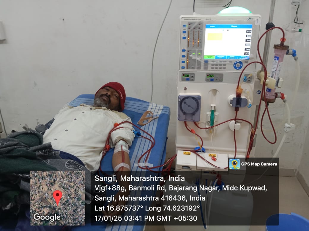
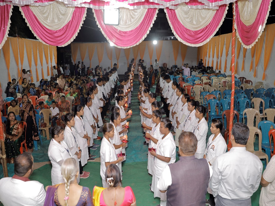
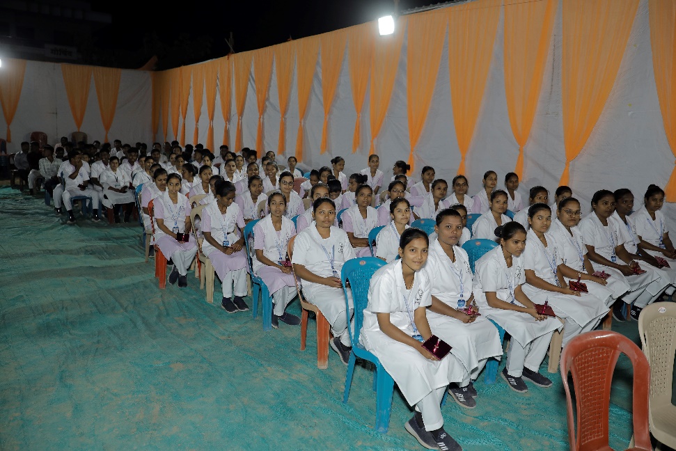
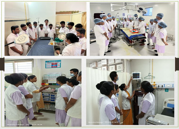
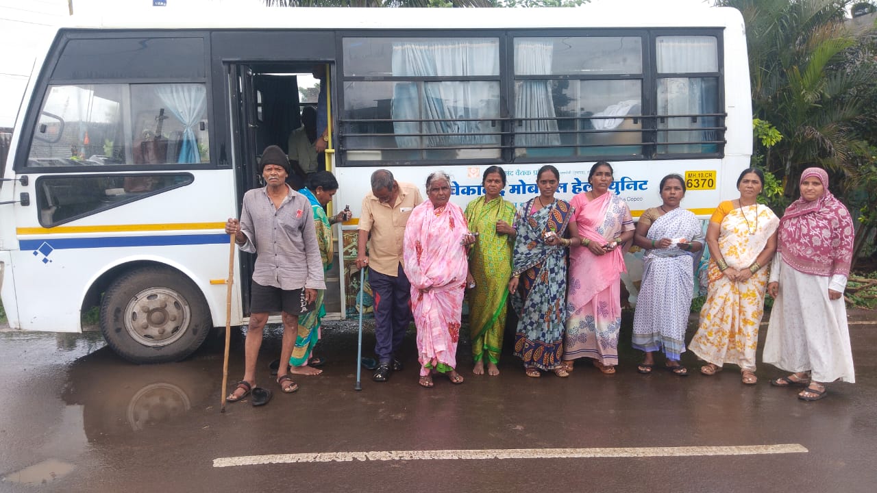
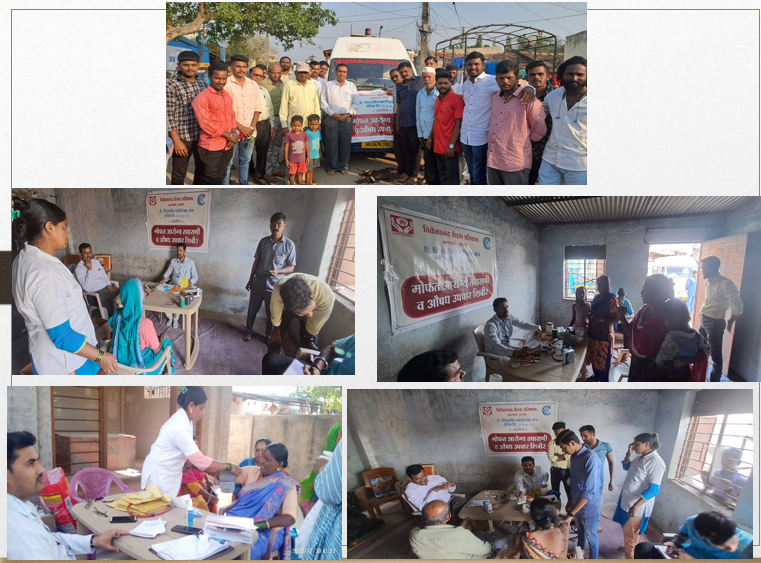
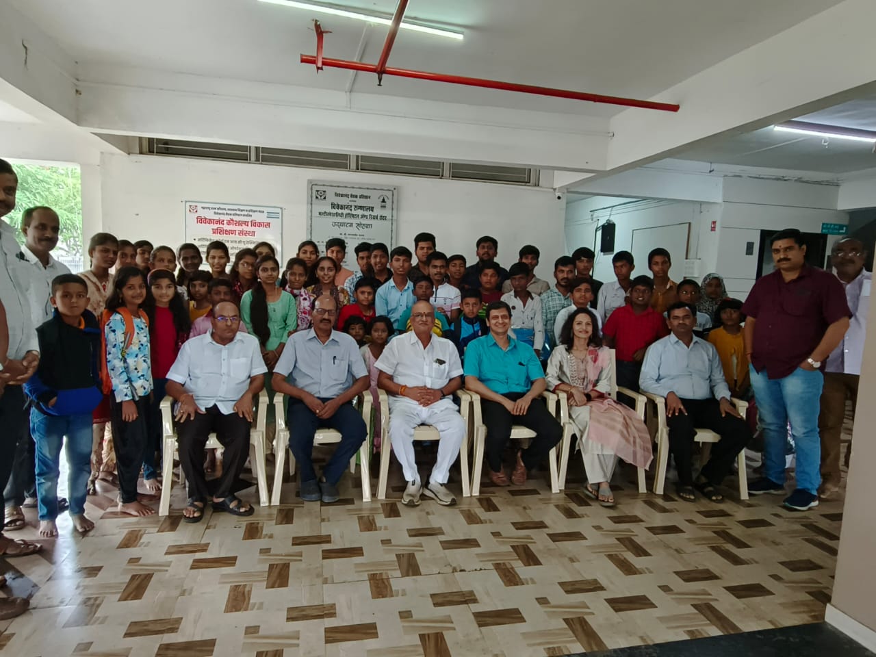
From the REC Ltd. MoA signing to hospital teams, community outreach, and clinical care — moments that frame the Cath Lab journey.
Latest state-of-the-art single-plane catheterization lab with flat-panel detector technology, digital imaging, and interventional vascular capability.
SINGLE PLANE CATHLAB
Technical Specifications of Latest state of art, single plane catheterization lab with flat panel detector technology digital imaging system with latest image processing and interventional vascular procedures, vascular angiography
| Clause | Specification |
|---|---|
| 1.0 | C-Arm Multi-directional, floor mounted |
| 1.1 | C arm should be latest design, for clear free floor space with head to toe covering of the patient. |
| 1.2 | It is possible to position the C arm on both side (Right or Left) of patient. |
| 1.3 | Arm design should allow sufficient space around the table during resuscitation and defibrillation. |
| 1.4 | Peripheral positions and vascular intervention should be available. |
| 1.5 | System should have facility for more than 10 user defined programs positions for C-arm. |
| 1.6 | Arc Depth : 823 mm +/-1 mm L = 0°: LAO 90° +/-2°, RAO 90° +/-2°, CRA 45° +/-2°, CAU 45° +/-2° L = 90°: LAO 45° +/-2°, RAO 45° +/-2°, CRA 45° +/-2°, CAU 45° +/-2° Orbital Speed : 10°/sec. to 12°/sec. Rotational Speed: 24°/sec. +/-2°/sec. L-Arm Rotation: +/-90° +/-2 L-Arm Movement speed: 12°/sec to 14°/sec FPD Movement: 300mm +/- 05mm FPD Up/Down speed : 10mm-40mm/sec |
| 1.7 | Contact based collision sensor at tube side. |
| 1.8 | Two level collision protection at detector side: Non-contact based collision sensor Contact based collision sensor |
| 1.9 | Patient Table |
| 2.0 | Floor mounted table with easy patient transport capability with longitudinal, transvers movement and motorized vertical movement. 3-Axis carbon fiber table top with minimum attenuation & electromagnetic lock. |
| 2.1 | Maximum patient weigh 200kg +100kg (additional force during resuscitation.) |
| 2.2 | Other Requirements :- (approximate dimensions) Table top Length: 2900mm +/- 10mm. Table Top Width: 465mm +/- 05mm Tabletop height from floor: Min. 790 mm - Max. 1040 mm +/-10 mm Motorized Vertical up-down movement (motorized): 250 mm +/-10 mm & speed 20-22 mm/sec Longitudinal travel :1200mm +/- 10mm Transversal travel :320mm +/-10mm |
| 2.3 | Accessories for table should include head fixing aids, mattress, catheterization arm support, drip stand. |
| 3.0 | X-Ray Generator |
| 3.1 | Generator is of latest technology with high frequency type with atleast100kW output power. |
| 3.2 | KV Range (CINE) 40-125KV |
| 3.3 | KV Range (Fluoroscopy) 40-125KV |
| 3.4 | Pulsed Exposure: upto 30 fps |
| 3.5 | High mA Fluoroscopy: 10-130mA |
| 3.6 | High mA Cine: Maximum 1000mA |
| 3.7 | Generator should be at least 40 Khz or more frequency & of indigenous make. |
| 3.8 | Should have Digital ADRC for fluoroscopy. |
| 4.0 | X-Ray Tube |
| 4.1 | X-Ray tube should have minimum of two focal spots with high cooling rate to ensure continuous operation capable of pulsed fluoroscopy. |
| 4.2 | The X-Ray tube has anode heat storage capacity of at least 3.0 MHU to run continuously for 6-8 hours without shutting off. Generator should be able to provide continuous fluoro power of at least 3000W or more |
| 4.3 | Cooling system is with oil/water cooling to ensure continuous operations |
| 4.4 | Anode heat cooling rate is as per radiation safety norms of AERB. |
| 4.5 | X-Ray tube with noise less operation with anode heat storage capacity to support long intervention procedures without interruption. |
| 4.6 | Collimator: Multi Beam Filters. Preview collimation. Parallel Lead Shutters (Long & Cross) with automatically adjust as per SID to reduce scatter dose. Half cardiac shutter |
| 5.0 | Radiation protection |
| 5.1 | The system have integrated automatic controlled X-Ray beam filtering with copper filters of various suitable sizes as per radiation safety norms of AERB. |
| 5.2 | The system has positioning of collimator blades radiation for radiation protection. |
| 5.3 | The system has monitoring and display of X-Ray dose during the patient examination. Dose Area Product (DAP) meter is provided to record the Dose given to the patient |
| 5.4 | System meet all national and international safety standards and comply with BARC/ AERB guidelines. |
| 5.5 | System offered latest radiation safety techniques. |
| 5.6 | Pre-programming for image setting for different operating Modes |
| 6.0 | Flat Panel Detector |
| 6.1 | An amorphous silicon receptor Flat panel detector with size of 20cm x 20cm |
| 6.2 | Pixel size not more than 194 microns. |
| 6.3 | Digital System with acquisition and processing in 1Kx1K matrix 16 bit digitization. |
| 6.4 | The DQE of detector should be 80 %. |
| 7.0 | Monitor/Display: |
| 7.1 | The monitor display system in examination room is ceiling suspended. |
| 7.2 | It is possible to position it on the left or right side of patient table. |
| 7.3 | Ceiling Mounted monitor stand with telescopic up/down travel & Straight travel. |
| 7.4 | Monitor at procedure Room 55" Colored (01 No) + 32" Colored (01 No) Monitor at Console Room 43"Colored (1 No) +21" Colored (1No) |
| 8.0 | Digital Imaging System & Archiving: |
| 8.1 | Image processing software with real time image capturing, storage, and display in 1kX1k format. Boosted fluoroscopy (CINE) at 30 FPS with real time recording on hard disk drive. |
| 8.2 | Multi Views on Single 55 inch monitor with 4k Resolution User selectable multi format 55" display. Different types of views like Fluoro /Cine, DSA, Roadmap, Stent View and ECG parameters on single display. Configurable multiple View mode and positions of viewer can be swapped by the user as per convenience. |
| 8.3 | Dose Saving Protocol: Real time graphical and numbered dose display with dose reduction suggestion in real time. |
| 8.4 | Multi Para Monitor integrated in Single 55 inch monitor along with Fluoro/Cine, DSA, Roadmap and stent view. |
| 8.5 | Up to 10 FPS image acquisition for DSA Both Subtracted & Un-subtracted Parallel display R-Mask Landmarking(Intensity based) Pixel Shift |
| 8.6 | Real time path map Both Subtracted & Un-subtracted Parallel display Roadmap clear Peakhold |
| 8.7 | Image Processing features: Real time noise reduction filter without any motion lag DRC, Contrast, Brightness, Sharpness, Noise reduction. Interactive digital Zoom and Pan Pre-programming for image setting for different operating Modes Image Inversion WW/WL level adjustments Image Flipping and Image Rotation Clockwise or Anti-clockwise. Live to Reference View on Single Monitor Cine Loop Play(Auto and Frame wise) Real time Image Flip(Horizontal/Vertical) Real Time Image Inversion Multi Ref. Image Storage |
| 8.8 | Stent View: It should enhances the view of placed stent. |
| 8.9 | Cardiac & Vascular CO2 procedure should be done |
| 8.10 | Detection of luminal obstructions in Coronary Artery vasculature. It quantifies the plaque in the artery. |
| 8.11 | QLVA for the quick, convenient and accurate quantification of left-ventricular volumes and wall motion from left ventriculogram. |
| 8.12 | Ultra-fast Preview collimator with SID map feature to minimize the radiation dose to the patient as well as to the user. |
| 8.13 | DAP dose integrated in software and total summary for Fluoro/Cine Saved. |
| 8.14 | Structured Dose report: DICOM structured report for Dose detail based on the dose delivered to the patient is provided. |
| 8.15 | Real Time 4 step Digital MAG. |
| 8.16 | DICOM 3.0 compatible Connectivity with DICOM workstation/PACS |
| 8.17 | 1TB SSD for faster storage (Minimum 1 lakh images can be stored). Additional 500 GB SSD Hard Disk for data backup. (Expandable as per the requirement). Post Fluoro Saving as per user need. |
| 8.19 | Stenosis measurement, Length Measurement |
| 8.20 | Rectangle, Ellipse, Line, polygon, Curve, Closed-Curve, Pointer, Freehand Point, Ruler, Protractor, (Angle Measurement), Text. |
| 8.21 | Multiple DICOM Nodes can be configured to transfer image data Single/Multiple Image Tagging to transfer into PACS/ Workstation |
| 8.22 | Auto image transfer can be done during procedure in background mode. |
| 8.23 | User can adjust the patient's orientation to suit the procedure which is being performed and to match their actual position on the table. |
| 8.24 | Different format of image saving like JPG, BMP, TIF, GIF. Multi-Language GUI Support |
| 9.0 | Hemodynamic system 3/5 lead ECG amplifier. Dual IBP available in MPM SPO2 measurement Respiration : available NIBP measurement Temperature : 2 channels : range 0 degree 50 degree Cel. |
| 10.0 | UPS: Suitable online UPS with 30 min battery backup for complete Cath Lab including cine and fluoroscopy. |
| 11.0 | Accessories to be supplied:- Lead Glass of 2mm lead equivalent (120cm x 100cm) ( as per national and international safety norms). Radiation Shield - ceiling and table mounted radiation protection ( as per national and international safety norms). Integrated two way communication system between control room and examination room. Ceiling suspended operational light 4Nos.good quality lead aprons 2Nos.Thyroid guards 2Nos. lead cap. 01No. Pressure injector of repute make. |
| 12.0 | Environmental factors A. The unit shall be capable of being work continuously in ambient temperature of 19°C - 21°C and relative humidity of 0% - 50%. B. Proper Shielding should have to be done by the concessionaire to minimum radiation leakage as per AERB and BARC regulations. |
| 13.0 | Power Supply A. Power input to be 400-440 V ( 3 Phase )/ 50 Hz as appropriate fitted with Indian plug. B. Reset Table over current breaker shall be fitted for protection. |
| 14.0 | Standard Safety and Training Cath and accessories should be as per National and International parameters. Electrical safety conforms to standards of electric safety IEC -60601-1 General Requirements. Manufacturer should have ISO certifications for quality standards. Shall comply AERB guidelines. The system must be US FDA/ European CE with Notified Body approved |
| 15.0 | Documentation USER manual in English Service Manual in English List of important spare parts and accessories with their part number and costing. Certificate of calibration and inspection the factory. |
| 16.0 | Other Requirement A Model should be latest. B. Comprehensive warranty for 1 year for the complete systems including x-ray tube. C. Quote comprehensive maintenance contract (CAMC) for complete systems including x-ray tube for additional 5 years after expiry of warranty. D. The Company should be ISO 13485 &ICMED13485 certified. E. The company should have valid import//manufacturing license from central licensing authority or state licensing authority of CDSCO for Medical devices. F. The company should have proven track record. |
| 17.0 | Note: X-ray Generator/ X ray tube/Flat panel detector/Hemodynamic Monitor / Software-out of these at least 3 parts should be mandatory manufactured by participating principal company. |
Reach our team for details on cardiac cathlab services, facilities, and how this CSR-supported initiative serves the community.